PRISM Clinical education hub
PRISM* brings perfusion, bacterial status, infection staging, structural correction, and longitudinal monitoring into one imaging-first workflow. This page helps clinicians understand how the algorithm works, how Kent Imaging technology supports each step, and where to find the evidence, cases, and tools needed for implementation.
*PRISM is a clinical framework developed and clinically vetted by practicing clinicians
Why PRISM matters
Chronic wounds, especially diabetic lower-extremity wounds, often stall because three modifiable barriers remain hidden at the bedside: inadequate perfusion, unrecognized bacterial burden, and biomechanical deformity. PRISM gives clinicians a way to separate those barriers and respond to each one with documented findings.
Clinical foundation
Evidence-based imaging workflow
Technology role
How the Snapshot Suite supports
PRISM stands for Perfusion Imaging, Regional Bacterial Status, Infection Staging, Structural Correction, and Maintenance & Monitoring. In plain language, it is a structured algorithm that helps wound care teams move from objective imaging findings to the next clinical decision without relying on visual impression alone. PRISM is published by a multidisciplinary panel of wound care, podiatric, and vascular specialists.

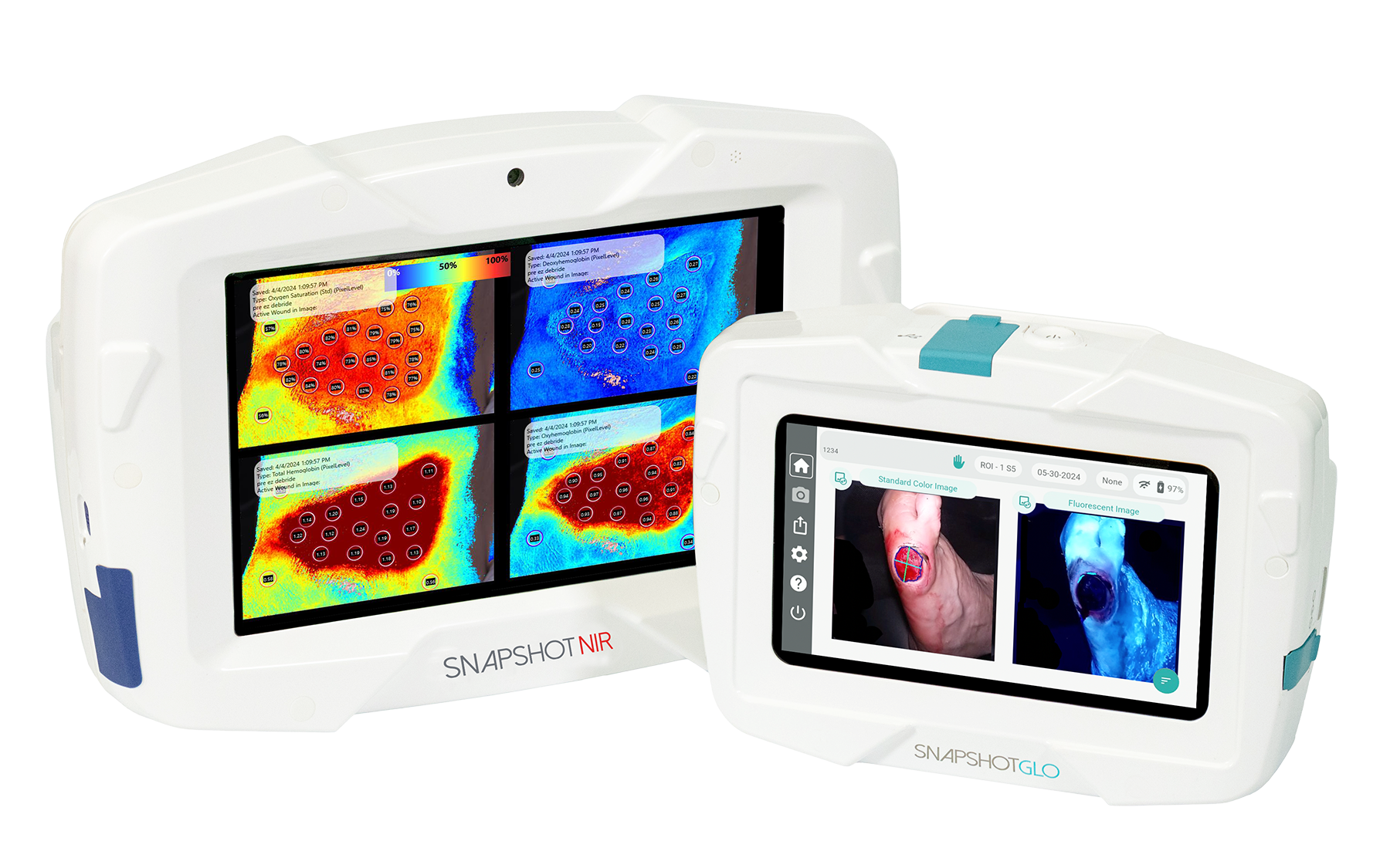
SnapshotNIR maps tissue oxygen saturation and related hemoglobin views so clinicians can see whether the wound environment can support healing before they escalate treatment.
SnapshotGLO reveals red and cyan fluorescence before wound manipulation, helping clinicians localize bacterial burden that may not be obvious on exam.
The algorithm ties those findings to infection staging, structural decisions, and follow-up intervals so care can stay consistent across providers and settings.
PRISM Summary: Objective imaging supports a repeatable clinical standard, which supports consistency of care, practitioner adoption, and stronger implementation of Snapshot technology within the wound workflow.
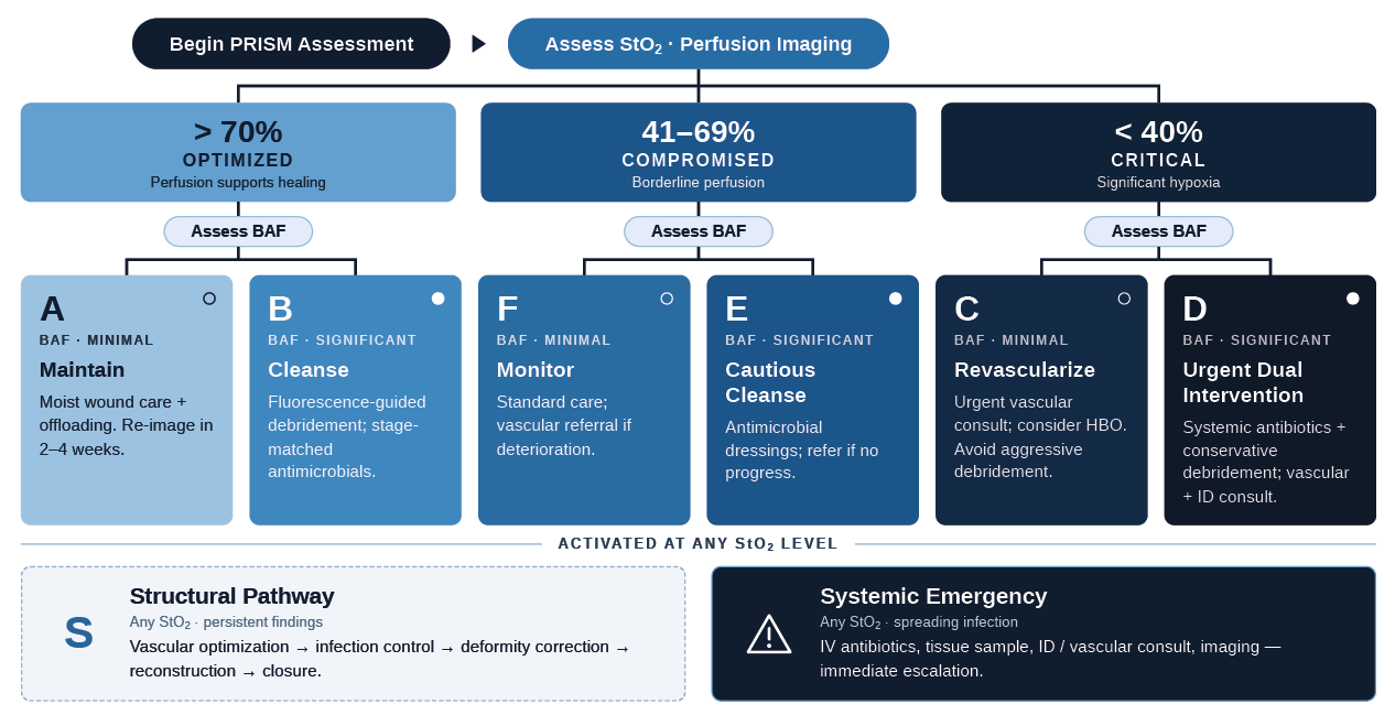
PRISM is designed as a sequence that can be repeated at each encounter. Perfusion Imaging, Regional Bacterial Status, and Infection Staging happen every visit. Structural Correction and Maintenance & Monitoring are activated according to what the imaging and clinical context show.
Use near-infrared spectroscopy to map StO2, HbO2, and DeOxyHb so perfusion risk is visible before the wound is manipulated.
Capture bacterial autofluorescence before hygiene to localize red and cyan fluorescence and document the baseline bacterial picture.
Translate fluorescence findings into staged, evidence-informed antimicrobial decisions that can be documented and trended over time.
Address deformity, recurrent pressure, or osteomyelitis-driven anatomy when wound closure will not hold without biomechanical correction.
Set an imaging cadence, reinforce offloading and education, and coordinate multidisciplinary follow-up to protect healing progress.
Interactive Walkthrough
PRISM is imaging-first by design. Capture NIRS before wound hygiene to assess perfusion. Capture BAF before manipulation to map bacterial burden. Stage infection using the fluorescence findings, then decide whether the case needs structural correction, closer monitoring, or urgent escalation.
1. Start with physiology
SnapshotNIR shows whether tissue oxygenation is adequate, in a vigilance zone, or significantly compromised.
2. Add bacterial context
SnapshotGLO identifies wound-bed, periwound, or spreading fluorescence so bacterial burden can be localized before cleansing or debridement.
3. Match action to stage
Use staged interventions that range from standard care to targeted antimicrobials, tissue sampling, or urgent infectious disease and vascular consultation.
4. Reassess and document
Repeat the sequence at follow-up visits so teams can confirm bacterial clearance, monitor perfusion trends, and standardize documentation across providers.
StO2 above 70% with minimal bacterial burden supports maintenance, offloading, and routine re-imaging.
StO2 above 70% with significant burden supports fluorescence-guided cleansing, debridement, and stage-matched antimicrobials.
StO2 41–69% with minimal burden calls for closer surveillance and trend-based vascular vigilance.
StO2 41–69% with significant burden supports cautious cleansing, antimicrobial dressings, and escalation if progress stalls.
StO2 below 40% with minimal burden shifts the priority to vascular evaluation and perfusion optimization.
StO2 below 40% with significant burden calls for infection control and vascular intervention in parallel.
Persistent burden with a structural driver activates the surgical and biomechanical pathway toward durable closure.
Spreading fluorescence with infection concern is an emergency pathway that increases urgency, sampling, imaging, and consultation.
PRISM is built for routine use at the point of care. The sequence supports wound specialists, podiatric and vascular surgeons, and multidisciplinary limb-preservation teams that need a common decision path across visits and care settings.
SnapshotNIR provides real-time tissue oxygen saturation, oxyhemoglobin, deoxyhemoglobin, and total hemoglobin views so clinicians can classify perfusion before they choose debridement intensity, vascular escalation, or follow-up cadence.
SnapshotGLO captures bacterial autofluorescence to show red fluorescence from general bacteria and cyan fluorescence associated with Pseudomonas, helping teams target debridement, document clearance, and support antimicrobial stewardship.
Future innovations from Kent will be developed with the PRISM algorithm at their core, extending standardized, imaging-driven wound assessment across new modalities and clinical applications.
Capture baseline clinical images and measurements
Document the wound, periwound, comorbidities, and recent interventions at the start of the encounter.
Perform NIRS imaging before wound manipulation
Use SnapshotNIR to assess StO2 and identify whether perfusion is adequate, intermediate, or significantly compromised.
Perform BAF imaging before hygiene or debridement
Use SnapshotGLO to localize bacterial burden and assign the next infection-staging step from wound-bed to spreading patterns.
Act on the combined findings
Match the imaging to vascular referral, targeted antimicrobials, conservative or aggressive debridement, or structural correction planning.
Repeat, trend, and coordinate follow-up
Re-image at the appropriate interval, document bacterial clearance and perfusion trend, and coordinate offloading, glycemic management, and multidisciplinary follow-up.
PRISM is strongest when clinicians can move from framework to proof. This hub is designed to hold the publication, clinical posters, real-world cases, expert commentary, downloadable tools, and follow-up education that support broader understanding and adoption. Materials will be added as they become available.
Click the links below to access resources that are already available.
Supporting evidence and practical steps needed to use PRISM consistently across sites of care.
PRISM starts before wound hygiene or debridement. Capture NIRS first to assess tissue oxygenation, then capture BAF before manipulation so bacterial burden is documented at baseline. That sequence gives the clinician an objective starting point for infection staging, escalation decisions, and follow-up planning at the same visit.
PRISM gives teams a shared order of operations and a shared language for what the images mean. When perfusion, bacterial status, infection stage, and structural triggers are documented the same way, it becomes easier to align treatment decisions across wound clinics, hospital services, and limb-preservation programs.
SnapshotNIR supports the perfusion decision by showing tissue oxygenation and related hemoglobin views at the bedside. SnapshotGLO supports the bacterial-status and infection-staging decisions by localizing red and cyan fluorescence before wound manipulation. Together they provide the physiologic inputs that PRISM uses to guide the next step.
No. PRISM is designed to support clinical judgment with repeatable, point-of-care imaging. It gives clinicians a structured way to document what they see, act earlier on hidden barriers to healing, and monitor whether the response is changing over time without presenting the technology as the decision-maker.
For partnership inquiries and clinician follow-up, please email clinical@kentimaging.com